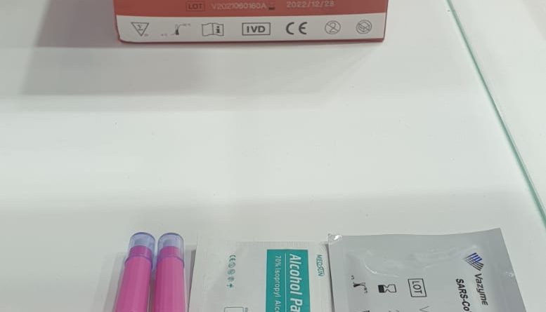
SARS-CoV-2 RBD Protein IgG Detection Kit (Colloidal Gold-Based)
The global outbreak and pandemic of the novel severe acute respiratory syndrome coronavirus 2 (SARS-CoV-2) have a significant impact on the global economy and human health. To reduce the spread of the disease, immunized mice generated 16 monoclonal antibodies (MAbs) against SARS-CoV-2 with the spike protein receptor-binding domain (RBD), which were expressed in Chinese hamster ovary cells ( CHO). A colloidal gold-based immunochromatographic strip with two McAbs was developed to detect the SARS-CoV-2 spike protein, which may play a potential role in vaccine quality control.
The strip is highly specific as it detects only the SARS-CoV-2 spike protein and does not show any non-specific reaction with syndrome coronavirus (SARS-CoV), Middle East respiratory syndrome coronavirus (MERS-CoV) and other coronaviruses and influenza viruses. . The strip detected the subunit vaccine in our laboratory with a spike protein detection limit of 62.5 ng/mL. This strip provides an effective method of monitoring vaccine quality by detecting the antigen content of the spike protein.
Introduction
Coronavirus disease 2019 (COVID-19) caused by the novel severe acute respiratory syndrome coronavirus 2 (SARS-CoV-2) infection is spreading to more than 200 countries/regions with 50 million infections and 1,263,844 deaths ( until November 12) according to statistics from the World Health Organization. Rapid antigen detection (RAD) tests play an important role in reducing the spread of the disease. On the one hand, RAD tests have the potential to be an important tool for the early diagnosis of SARS-CoV-2, especially in cases where molecular methods are limited.
On the other hand, RAD can monitor the amount of SARS-CoV-2 spike protein available in vaccines. Although RT-PCR and viral culture are the current gold standards in the diagnosis of SARS-CoV-2 infection, several factors, such as trained personnel, the need for special equipment, and the price of the reagent, limit their use. of these time-consuming molecular techniques. Therefore, investigation of different immunochromatographic strips for the detection of COVID-19 is encouraged.
SARS-CoV-2 is a single-stranded positive-stranded RNA virus. Its genome encodes four structural proteins: spike protein (S), small protein (E), matrix protein (M), and nucleocapsid protein (N). The Spike protein is a type I fusion protein that forms trimers on the surface of viral particles. It consists of two subunits, S1 and S2: S1 mainly contains the receptor-binding domain (RBD), which is responsible for recognizing cellular receptors, and S2 is responsible for membrane fusion. Angiotensin-converting enzyme 2 (ACE2) is used to enter target cells as a receptor.
Therefore, the RBD of the spike protein determines the infectivity of the virus and its propagation in the host. Although the immunochromatographic strips developed by some researchers use antibodies against the N protein of SARS-CoV-2. Most of the vaccines and antibody therapies in development are directed against the RBD spike protein of SARS-CoV-2, as the protein is the main antigen that induces a protective immune response.
On April 7, 2020, a large-scale case study published by the CDC in the United States estimated that the Basic Reproduction Number (R0) of SARS-CoV-2 was 5.7, which means that the ability to SARS-CoV-2 transmission is much higher than that of SARS-CoV (R0: 0.85-3). The world urgently needs an effective and safe vaccine, which will play a decisive role in controlling the global epidemic. According to the draft vaccine candidates published by the WHO on August 27, 2020, there are 143 vaccines worldwide in preclinical trials, 33 vaccines have entered clinical trials, of which 8 vaccines have entered phase III clinical trials.
Recently, the vaccines produced in China have been fully commercialized. Vaccines being investigated include widely used traditional vaccines, namely inactivated or attenuated vaccines, genetically modified recombinant subunit vaccines, adenovirus vector vaccines, recombinant virus vector vaccines, and newer vaccines that have not been approved for similar vaccines. , mainly those that include ribose Nucleic acid (RNA) vaccines and deoxyribonucleic acid (DNA) vaccines. Among these vaccines, the leading protein-based vaccines occupy a certain number.
Materials and methods
- Statements of Ethics and Biosafety
The experimental research protocol for monoclonal antibody production in mice was approved by the Key Laboratory of Animal Immunology, Henan Academy of Agricultural Sciences, China, in accordance with its policies and procedures.
- Expression and purification of Spike RBD protein
The sequence used for the protein was based on the genomic sequence of the first isolate, Wuhan-Hu-1, which was published on January 10, 2020 (peak protein residues 132–537, GenBank: MN908947.3). The signal peptide sequence of the SARS-CoV-2 RBD spike protein (spike protein 1–18 residues) was added to the N-terminus of the protein for protein secretion, and a Hexa-His tag was added to the C-terminus of the protein for further purification. These constructs were synthesized by Sangon Biotech (Shanghai, China).
These constructs were cloned into the pcDNA3.1 (+) vector and transiently transfected into CHO cells when the cell density was 6 × 10 6 cells/mL. After 3 days, cells were harvested and centrifuged for 10 min at 10,000 × g, 4 °C. The supernatant was filtered with a 0.22 µm Stericup filter and purified by Ni affinity chromatography on a 5 ml HisTrap™ excel column (GE Healthcare) in a buffer consisting of 20 mM PBS (pH 7.2). The protein size and purity of the elution peak were analyzed by SDS-PAGE and the protein concentration was determined with the BCA protein concentration determination kit.
- Generation of Monoclonal Antibodies
Monoclonal antibodies against SARS-CoV-2 were developed following a standard procedure. Briefly, 6-week-old female BALB/c mice (n = 5) were immunized with the RBD spike protein produced in this study at an immunization dose of 0, 20, 50, 100, and 200 μg each mouse in adjuvant. Freud, respectively. Mice were immunized every 2 weeks. Two weeks after the third immunization, blood was collected to determine mouse serum titer by ELISA.
The mouse with the highest titer was selected for superimmunization based on the amount of protein in the first immunization. Cell fusion was performed 3 days after superimmunization. Briefly, the mouse spleen was aseptically taken for grinding and then fused with Sp2/0 myeloma cells in a 1:2 ratio. Hybridoma cells were screened by ELISA and cloned by the limiting dilution method. Ascites fluids from positive hybridomas were produced in mice.
- Preparation of colloidal gold and gold-labelled monoclonal antibodies
Colloidal gold was prepared by the trisodium citrate method. Briefly, 1 mL of 1% chloroauric acid was added to the Erlenmeyer flask with 99 mL of bidistilled water stirring and heating, followed by the rapid addition of 1.6 mL of 1% trisodium citrate solution with rapid stirring. The mixture was boiled for another 5 min and gradually boiled until the colour gradually changes from light yellow to deep red and no longer changes colour. The colloidal gold solution was cooled to room temperature and then stored at 4 °C.
MAbs were centrifuged at 12,000 × g for 5 min and incubated with a colloidal gold solution for 30 min. 10% bovine serum albumin (BSA) was then added to the colloidal gold conjugate and incubated for 10 min. The mixture was then centrifuged at 12,000 × g, 4 °C for 30 min to remove any unbound antibody. The pellet was resuspended in boric acid buffer containing 1% BSA.

